*for hardware setup and calibration, please review the Doppler Flow Velocity System user manual.
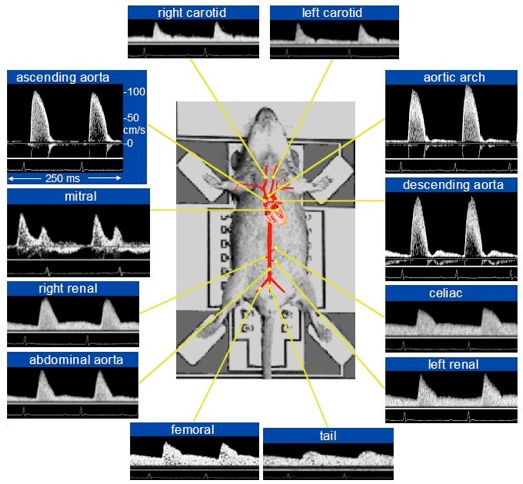
The subject is anesthetized with Isoflurane, an injectable cocktail, or other anesthetic agents depending on the desired type of study. The anesthetized animal is then placed in the supine position with its four limbs taped to the non-invasive electrodes on the Rodent Surgical Monitor. The Rodent Surgical Monitor captures, processes and filters the ECG signals and outputs the filtered ECG waveform to the Doppler Signal Digitizer. Blood flow velocity signals may be captured from the aortic outlet or mitral inlet by positioning the tip of a pulsed Doppler tubing mounted probe just below the sternum. A 10MHz probe is typically used for measuring flow velocity signals from deeper/farther sites of blood flow, while a 20MHz probe is typically used for shallower sites of blood flow. Blood flow velocities in peripheral arteries can be obtained using a 20MHz probe.
The 10 & 20 MHz pulsed Doppler transducers are plugged into the 10MHz & 20MHz receptacles on the either channel A or channel B of the Pulse Doppler Transceiver which generates in-phase and quadrature demodulated audio signals that are sampled and digitized by the Doppler Signal Digitizer. In-phase and quadrature demodulated Doppler audio signals generated by other equivalent systems can also be input into the Doppler Flow Velocity System at the Doppler Signal Digitizer stage and displayed on the Doppler Workstation.
Leave a Reply